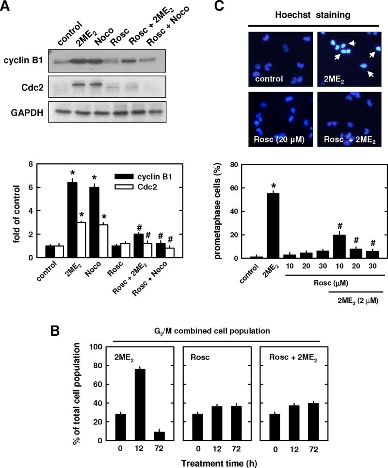
Fig. 4. Effect of roscorvitine on 2ME2-induced prometaphase arrest.
(A and B) MCF-7 cells were pre-treated for 2 h with roscovitine (20 μM) and then further treated with 2 μM 2ME2 or 250 nM nocodazole for additional 12 h. Total cell lysates were analyzed by Western blotting for cyclin B1 and Cdc2 protein levels (A, upper panel). The relative protein levels of cyclin B1 and Cdc2 are calculated according to their densitometry readings, which are normalized to GAPDH protein level (A, lower panel). (B) Cells were pre-treated for 2 h with roscovitine (20 μM) and then treated in combination with 2 μM 2ME2 for additional 12 or 72 h. Cells were harvested and analyzed using flow cytometry. Each bar is the mean ± S.D. from triplicate measurements. (C, upper panel) Gross nuclear morphological changes in cells treated with a representative concentration (20 μM) of roscovitine. The nuclear morphology was examined under fluorescence microscopy (× 200 magnification) after the cells were stained with Hoechst-33342. The arrow points to a prometaphase-arrested cell. (C, lower panel) Quantitation of prometaphase cells based on counting 200 nuclei in each treatment group. * P < 0.05 versus vehicle control; #P < 0.05 versus 2ME2 or nocodazole treatment.

