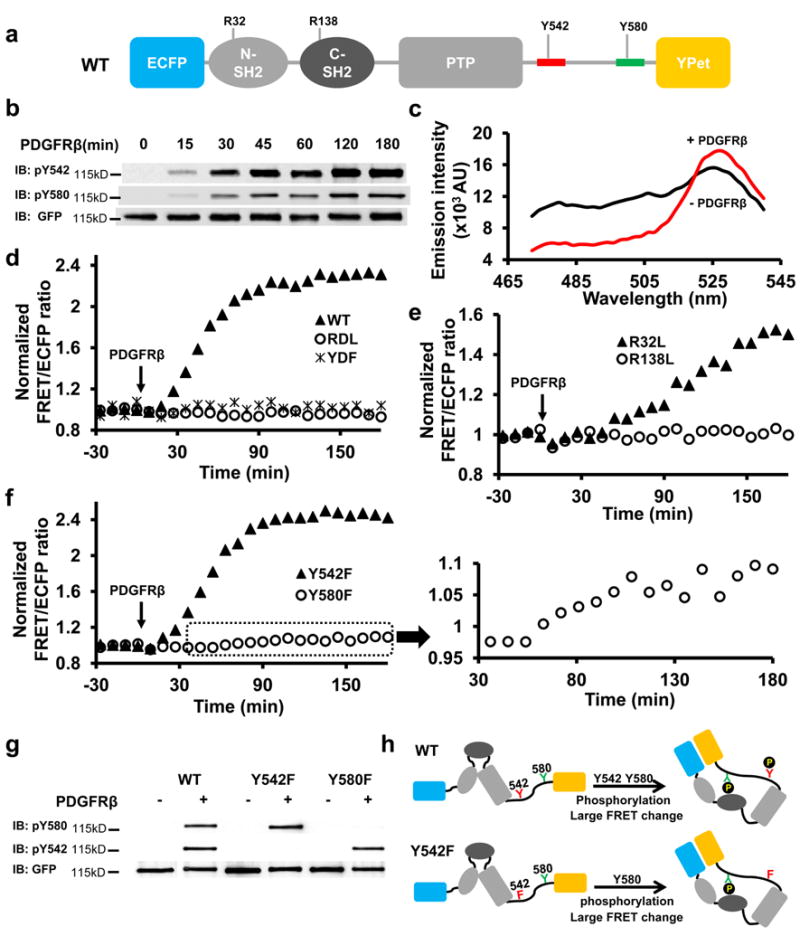
Figure 1. cis-Interactions of the Shp2 reporters in vitro.

(a) Schematic drawing of the WT Shp2 reporter, with mutation sites as indicated. Y542 and Y580 sequences are colored in red and green, respectively. (b) The phosphorylation of WT reporter upon PDGFRβ incubation in vitro(refer to Supplementary Figure S6a for the whole blots). (c) The emission spectrum change of the WT reporter before (black) and after (red) PDGFRβ incubation. (d-f) The ratio time courses of WT reporter and its mutants as indicated. (f) inset shows the time course of zoom-in ratios of Y580F reporter upon PDGFRβ incubation. (g) The tyrosine phosphorylation of Y580 and Y542 of the WT reporter, its Y542F or Y580F mutant in vitro before and after PDGFRβ incubation. (h) The models depict the cis-interactions and FRET changes of the WT and Y542F reporters upon phosphorylation in vitro.
