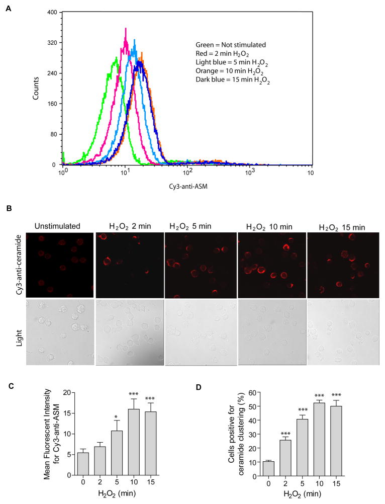
Fig. 1. Oxidative stress induces ASM externalization and formation of ceramide-enriched membrane platforms.
(A) Oxidative stress by H2O2 (1 mM) induces a translocation of ASM onto the cell surface. Surface ASM was detected by flow cytometry analysis of cells incubated with Cy3-labeled goat anti-ASM antibodies. Shown is a representative flow cytometry analysis from four independent experiments. (B) H2O2 (1 mM)-induced ASM translocation correlates with rapid clustering of ceramide in the membrane and formation of ceramide-enriched membrane platforms. Representative confocal fluorescence microscopy and light images from three independent experiments are shown. (C) Displayed are the summarized data showing the mean fluorescence intensity for Cy3-anti-ASM staining from four independent experiments. (D) The quantitative analysis of the data from four independent experiments, which each included analysis of 200 cells/time point, shows the formation of ceramide-enriched membrane platforms in a large proportion of the cells. Displayed is the percentage of cells with a ceramide-enriched membrane platform at indicated time points. Panel C and D show the mean ± S.D. Significant differences between treated and non-treated controls were determined by t-test and are indicated by *** (P<0.001, t-test) or * (P<0.05, t-test), respectively.

