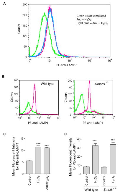
Fig. 5. Effect of ASM inhibition or deficiency on H2O2-induced lysosomal exocytosis.
(A) Shown is a representative flow cytometry analysis diagram using PE-conjugated goat anti-LAMP1 antibodies in Jurkat cells with or without ASM inhibitor amitriptyline (10 μM) (n = 4). (B) Spleen T cells were isolated from wild-type and Asm deficient (Smpd1−/−) mice, stimulated without (green) or with H2O2 (red) and stained with PE-conjugated goat anti-LAMP1 antibodies. Shown are representative flow cytometry analyses from 3 independent experiments. Summarized data from 3 independent studies show the mean fluorescence intensity for PE-anti-LAMP1 staining in Jurkat cells (C) or spleen T cells (D). Displayed is the mean ± S.D. Significant differences between treated and non-treated controls were determined by t-test and are indicated by *** (P<0.001, t-test).

