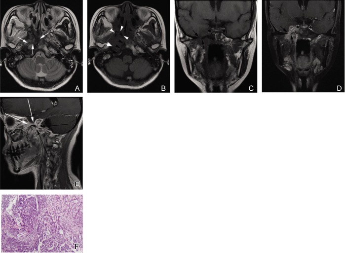
Figure 3. Solid subtype of NACC with a type 2b imaging pattern in a 25-year-old woman.
A, an axial T2W fast spin-echo image shows an irregular lesion with unclear margin in the right parapharyngeal space and right part of the skull base. The signal intensity of the lesion is heterogeneous and slightly hyperintense with hypointense separations (white arrows). B, an axial T1W fast spin-echo image shows the signal intensity of the lesion is similar to that of muscles. Absence of the normal hyperintense signal in the right pterygoid process (white arrowheads), the right petrous apex (curved white arrow), and the right part of the clivus (black arrowheads) indicates tumor invasion in the skull base. C and D, coronal T1W fast spin-echo and contrast-enhanced, fat-suppressed T1W images show that the tumor is localized in the right parapharyngeal space. The degree of enhancement is similar to that of normal mucosa. The tumor extends into the right cavernous sinus through the enlarged foramen ovale (black arrows). E, sagittal contrast-enhanced T1W spin-echo image shows the thickened mandibular nerve (long black arrows) and maxillary nerve (long white arrows). F, photomicrograph of a biopsy specimen shows the highly cellular, solid pattern of NACC (HE ×40).

