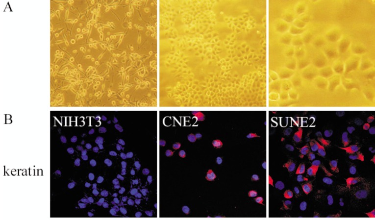
Figure 1. The morphology and keratin staining of the human nasopharyngeal carcinoma (NPC) cell line SUNE2.
A, SUNE2 cells grown in keratinocyte/serum-free (KSF) medium (×100, left panel) show feelers; SUNE2 cells grown in RPMI-1640 medium (×100, middle panel; ×400, right panel) firmly stick to the plate. B, SUNE2 cells are immunostained red in cytoplasm with anti-keratin, indicating their epithelial origin. NIH3T3 cells were used as a negative control, and CNE2 cells were used as a positive control.

