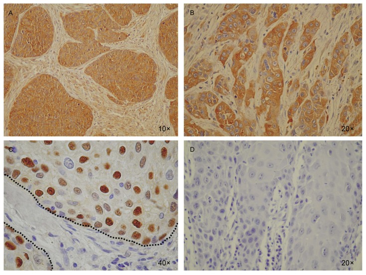
Figure 4. Id2, NF-κB, and Cyclin D1 are highly expressed in human SCC tissue sections.
A, the expression of Id2 was abundant in SCC cells (brown lumps) but not in non-cancerous cells (spotty in the normal tissue between SCC lumps). B, NF-κB showed a similar expression pattern as Id2 in human head and neck SCC specimens. C, Cyclin D1 expression was observed in the nuclei in a majority of SCC cells but was not detectable in the normal tissue between SCC lumps (between dashed lines). D, a representative immunohistochemistry control for A, B, and C.

