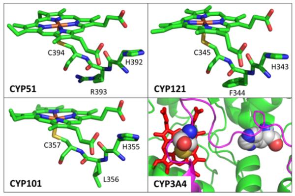
Figure 7.
Crystal structure of CYP51 (PDB#1EA1), CYP121(PDB#2IJ7), CYP101(PDB#2CPP) and CYP3A4(PDB#3NXU). The HXC motif in CYP51, CYP121 and CYP101 are highlighted. The right bottom panel shows the loop (magenta) of the heme proximal pocket in CYP3A4. The C442 and H402 residues are shown with a space filling model. There is no HXC motif in CYP3A4.

