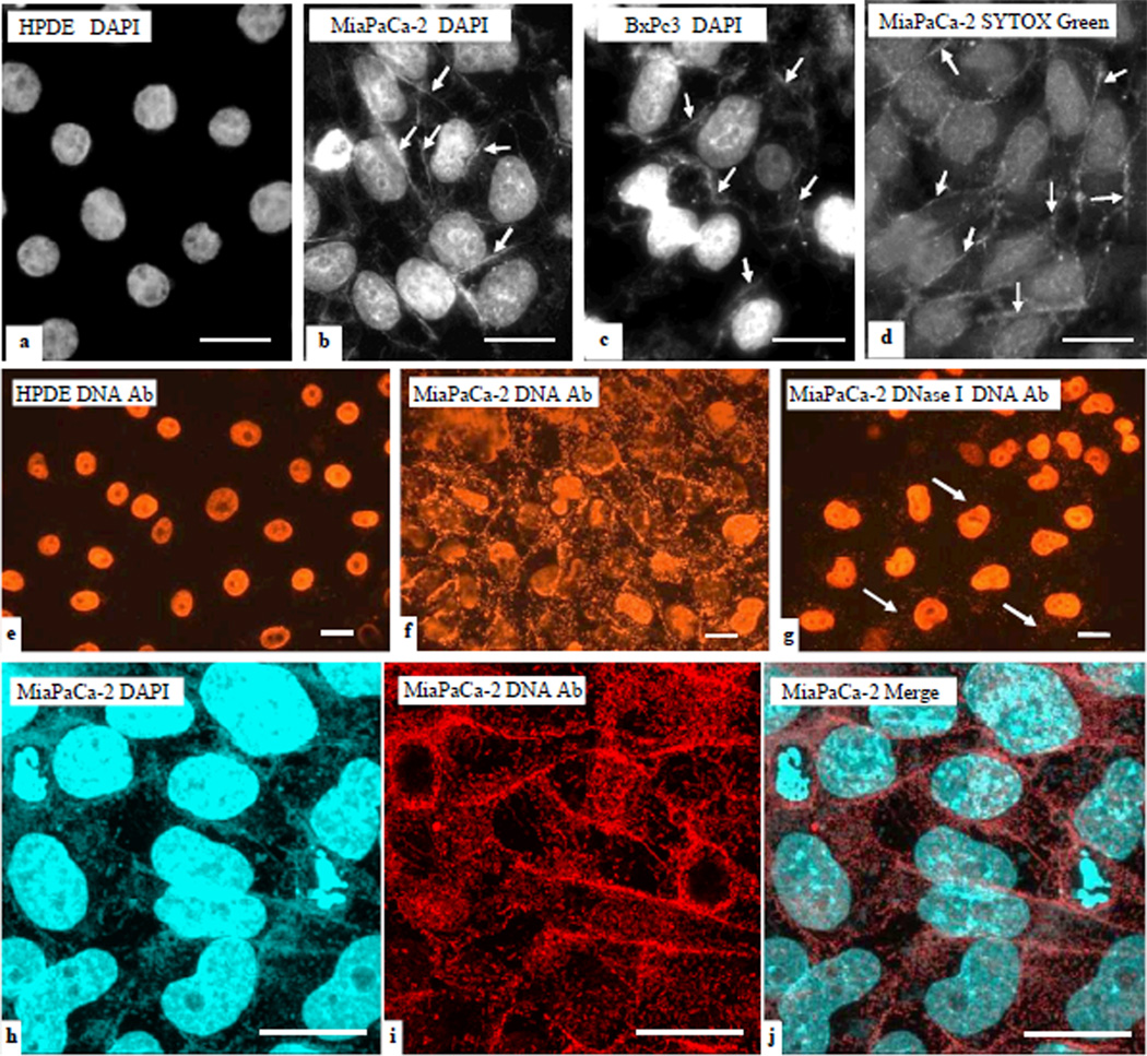
Figure 1. exDNA associated with pancreatic cancer cells, but not with normal pancreas cells.
Immortalized normal human pancreatic duct epithelial (HPDE), and human pancreatic cancer cell lines BxPC3 and MiaPaCa-2 cells were cultured on cover slips, and then fixed and stained with fluorescent DNA dyes DAPI (blue in a–c) and SYTOX Green (green in d). exDNA appears as fibrous materials (arrows). Bar = 20 µm. Immunofluoerescent stain (IF) using DNA antibody was conducted on HPDE, MiaPaCa-2, and BxPc3 cells. e. DNA antibody reacts to nuclei DNA in HPDE cells. f. DNA antibody reacts to exDNA and nuclei DNA in MiaPaCa-2 cells. g. MiaPaCa-2 cells were subjected to DNase I treatment before IF. e–g, images were taken under the regular fluorescent microscope. h–j, MiaPaCa-2 cells were double stained with DAPI and DNA antibody and images were taken under the confocal microscope. DAPI stain was done after DNA antibody. Cells in h–j were not permeabilized before staining whereas cells in e–g were permeabilized. Bar = 20 µm

