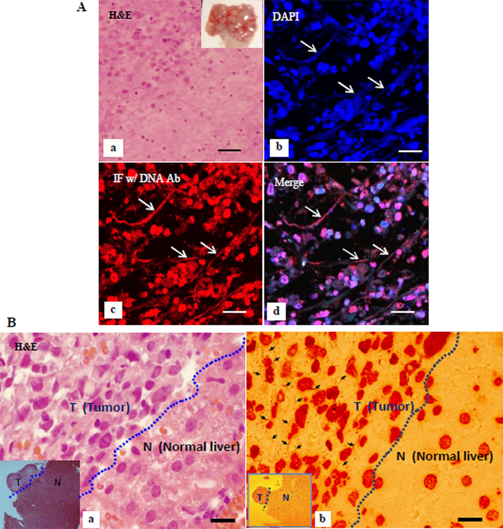
Figure 2. exDNA detected in vivo in mouse tissues.
A, pancreatic cancer metastasized to the diaphragm in a xenografted mouse. a, H&E of a tumor growing diaphragm (inset) section. b–d, double staining of a tumor growing diaphragm section with DAPI and imunofluorescent stain (IF) with DNA antibody, imaged under a confocal microscope. b, DAPI stain, c, IF with DNA antibody, and d, merge image of b and d. Arrows point to exDNA. Bar = 40um. B, exDNA detected in the metastasized tumor. IHC using DNA antibody revealed abundant exDNA (b, arrows) in a pancreatic cancer metastasized to liver tissue harvested from an orthotopically xenografted mouse. a, the corresponding H&E of IHC (b). No exDNA were observed in the normal liver tissue. Insets are lower magnification image using a 5× objective lens (T-tumor, L-liver). Bar = 20 µm.

