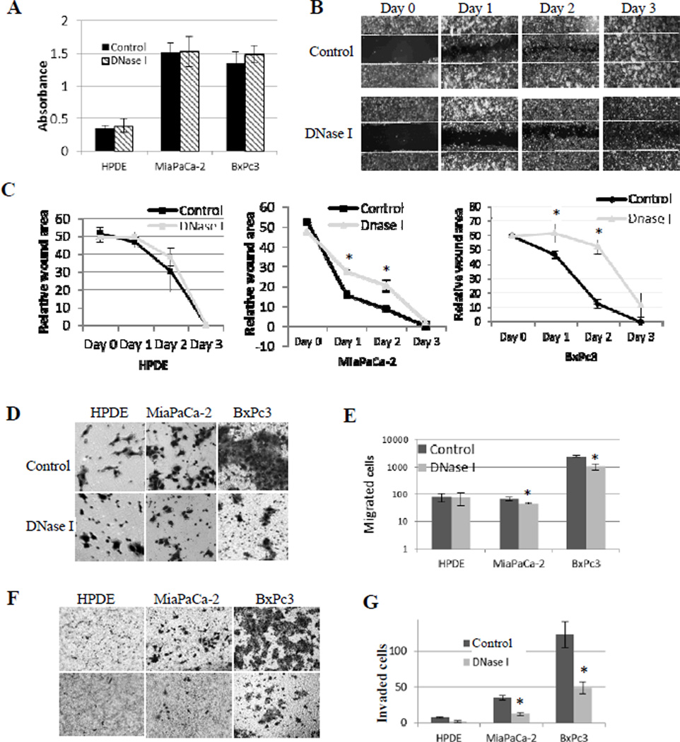
Figure 3. exDNA affects cell migration and invasion, but not cell growth in pancreatic cancer cell lines.
A, Cell proliferation was assayed by MTT method and the absorbance was read at 595 nm. Absorbance value positively correlated with cell numbers. Three triplicates sample in each plate per experiment. The results were reported as mean value of three experiments. B, Wounded areas were measured for 3 days after scratching the monolayer of cells in three cell lines. Images are representative of the wound-healing by MiaPaCa-2 cells. C, DNase I delays the wound healing in cancer cell lines, but not the HPDE normal cell line. Charts are plots of wounded areas of three cell lines measured through ImageJ of three random areas in three days. Data from three repeated experiments are presented as means ± SD, (*, p < 0.05, n=9). D, representative photographs of transwell-migration assay. Cells were seeded in transwells in the absence (control) and presence of DNase I (15 unites/well); after 24 hours, cells migrated to the basal side of the transwell membranes were stained with 0.5% Crystal Violet and 7 random views were counted. E, bar graph shows migrated cell numbers in normal pancreas HPDE cell line and two cancer cell lines. Data from three repeated experiments are presented as means ± SD, (*, p < 0.05, n=21). F, cell invasion setting is the same as for cell migration in D, except for transwell membranes were coated with Matrigel. G, bar graph shows numbers of cells invading through Matrigel in normal pancreas HPDE cell line and two cancer cell lines. Data from three repeated experiments are presented as means ± SD, (*, p < 0.01, n=21).

