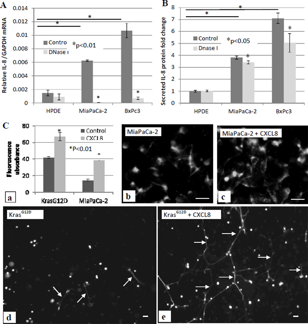
Figure 6. Feedback loop between exDNA and CXCL8.
Normal pancreatic HPDE cells, pancreatic cancer BxPc3 cells and MiaPaCa-2 cells were treated with DNase I. 500µl of 105 cells/well were seeded in absence and presence of DNase I (3 units/100µl) in a 24 well plate. 48 hrs after treatment, cells were harvested for total RNA isolation and qPCR to measure IL-8 mRNA. Relative fold changes of IL-8 mRNA level were normalized to GAPDH mRNA (A), and media were collected for ELISA assay to examine secreted IL-8 protein level. Fold changes of IL-8 protein level were against the level of IL-8 secreted from HPDE cells (B). C, MiaPaCa-2 and HPDE-KRASG12D cells were treated with 80 ng/ml exogenous CXCL8 for 18 hrs, then cells were stained with Sytox Green. exDNA were quantitatively measured (a) and were microscopically observed (b–e). Data from three repeated experiments are presented as means ± SD.

