
Figure 1.
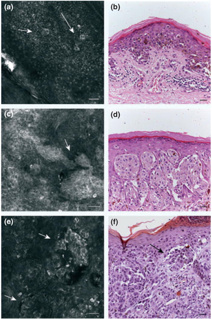
Confocal images of melanoma. (a) confocal image (500 × 500 μm) at epidermal level showing the presence of roundish pagetoid cells (arrows). (b) H&E stained section (bar = 50 μm). Original magnification 20×. Abundant pagetoid cells in a melanoma. (c) confocal image (500 × 500 μm) at dermo-epidermal junction: junctional nests showing single atypical cells and in contiguity with basal layer. (d) H&E stained section (bar = 50 μm). Original magnification 20× presence of junctional atypical clusters in a melanoma. (e) confocal image (500 × 500 μm) at dermal level: sparse nests (f) H&E stained section (bar = 50 μm). Original magnification 20×. Corresponding melanocytic proliferation forming discohesive nests in an invasive melanoma.