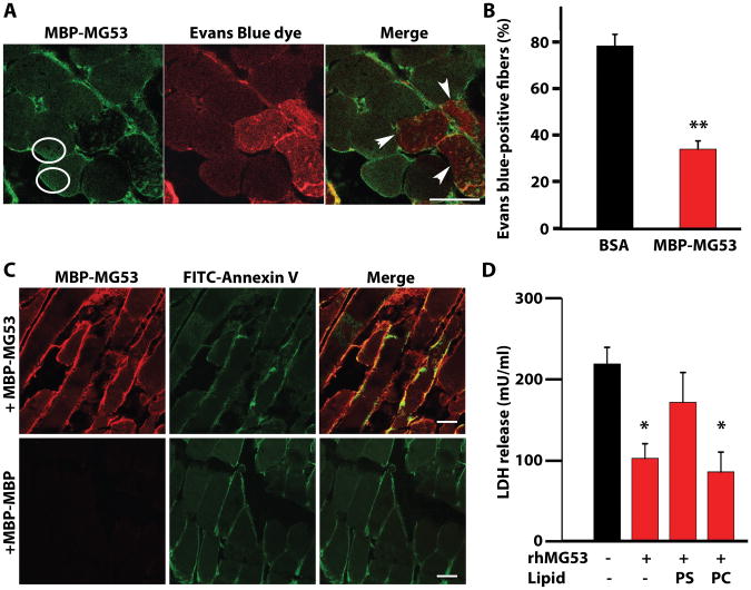
Figure 5. Cell membrane resealing mediated by rhMG53 requires binding to phosphatidylserine (PS).
(A) Mouse gastrocnemius muscles were co-injected with CTX, Evans blue dye, and either MBP-MG53 (20 μg/mL) or BSA. Sections were immunostained for MBP-MG53 and overlaid with images of Evans blue dye fluorescence. Circles surround areas of increased MBP-MG53 signal inside of muscle fibers. Arrows indicate fibers that lack MBP-MG53 decoration and contain Evans Blue dye. Scale bar, 20 μm. (B) Percentage of total muscle fibers displaying entry of Evans blue dye. Data are means ± SEM (n=24 sections) **P < 0.01 by ANOVA. (C) Gastrocnemius muscles of wild-type mice were co-injected with CTX, MBP-MG53, and FITC-labeled Annexin V, which binds to PS. Cryosections were stained for MBP-MG53 and examined by confocal microcopy. Control mice were injected with MBP-MBP instead of MBP-MG53. Images are representative of 4-6 sections from three different experimental animals per group. Scale bars, 20 μm. (D) HEK293 cells were treated with rhMG53 and damaged by glass microbeads before measuring LDH release. Cells were simultaneously treated with PS or phosphatidylcholine (PC) to check competition with rhMG53 function. Data are means ± SEM (n=6-8 independent assays with the condition performed in triplicate in each experiment). *P < 0.05 versus control without lipids or protein, ANOVA.

