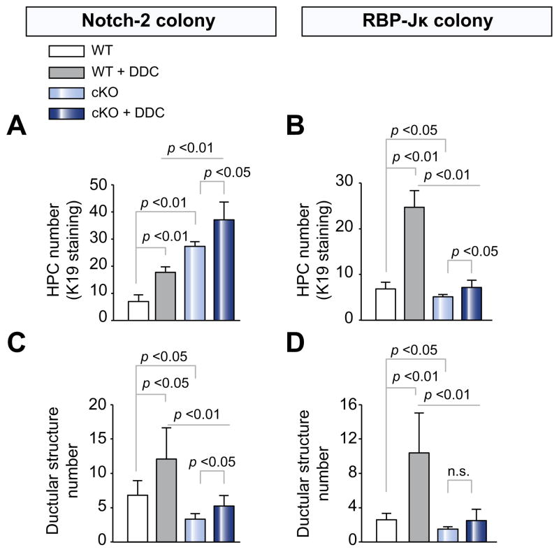
Fig. 3. Biliary progenitor cells and ductular structure quantification in WT, Notch-2-cKO, and RBP-JK-cKO mice after treatment with DDC.
Paraffin sections from Notch-2-cKO, RBP-Jk-cKO mice and their respective WT littermates, untreated or treated with DDC, were stained for K19. As shown in the bar graph, (A) Notch-2-cKO mice showed a significant increase in the number of HPCs after DDC damage. (B) Conversely, RBP-Jκ-cKO mice exhibited a significant reduction in the number of HPCs after treatment with DDC. On the other hand, both (C) Notch-2-cKO mice and (D) RBP-Jκ-cKO mice showed a significant decrease in the number of K19+ve ductular structures after DDC damage. (Data represent average ± SD, n = 4–8 mice per group; p values are reported). n.s., not significant.

