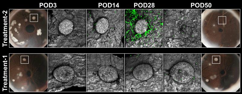
Fig. 6. Intraocular islet transplantation provides an experimental platform for screening new therapeutics longitudinally.
Serial photos and high-resolution confocal micrographs acquired on POD3, POD14, POD28, and POD50 of allogeneic pancreatic islets transplanted into the anterior chamber of the eye of mice expressing GFP in T cells. The mice were subjected to different experimental treatments administered systemically. Treatment-1 was with a proprietary lymphocyte-depleting agent and Treatment-2 was with an anti-chemokine agent. Digital images of the transplanted eyes on POD3 show individual islet grafts that were followed longitudinally using non-invasive in vivo imaging. Treatment efficacy was assessed based on islet infiltration by GFP-labeled T cells (green) and on islet structural integrity and survival in each treatment group. Islet grafts were visualized by laser backscatter (reflection; grey). These data demonstrate the utility of intraocular islet transplantation as a non-invasive drug screening tool during long-term in vivo studies.

