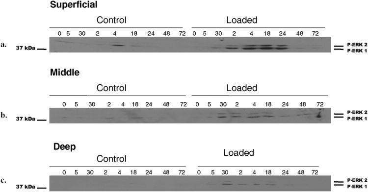
Fig. 1. Effect of mechanical compression on ERK 1/2 pathway activation.

Cartilage explant discs from the superficial, middle, and deep zones were mechanically compressed to 40% strain based on the post-equilibration explant thickness following a 48-72 hour equilibration in culture medium. A strain rate of 1sec- was utilized. The load was held for 5 seconds. Compression was performed in the presence of culture medium. Control explants were placed in a parallel non-communicating well within the loading device. Following compression, explants were processed as described in Experimental Procedures and analyzed using standard Western blot technique for ERK 1/2 phosphorylation using a phospho-ERK 1/2 specific antibody (a-c). Each band represents four individually compressed and pooled explants.
