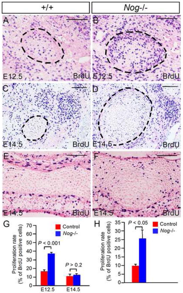
Figure 7. Lack of Noggin leads to elevated cell proliferation rate in Meckel’s cartilage.

(A–D) Coronal sections show BrdU labeled cells in control and Nog−/− Meckel’s cartilage at E12.5 and E14.5. (E, F) Coronal sections show BrdU labeled cells in presphehoid of E14.5 control (E) and mutant F). (G) Statistical analysis shows significantly increased cell proliferation rate in Nog−/− Meckel’s cartilage at E12.5 (P < 0.001) but not at E14.5 (P > 0.2) as compared to controls. (H) Statistical analysis shows significantly increased cell proliferation rate (P < 0.05) in the presphenoid in E14.5 Nog−/− embryo as compared to that in control. Standard errors are indicated. Scale bar = 100-μm.
