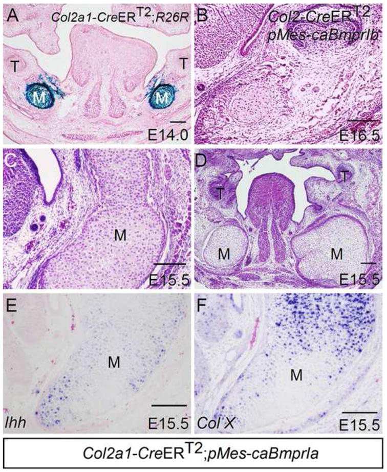
Figure 8. Activation of BMPRIa-mediated BMP signaling in chondrocyte-lineage resembles Nog−/− Meckel’s cartilage phenotype.

(A) X-gal staining shows Cre activity in chondrocytes of Meckel’s cartilage of an E14.0 Col2a1-CreERT2;R26R embryo that received an administration of tamoxifen at E12.5. (B) A coronal section of an E16.5 Col2a1-CreERT2;pMes-caBmprIb embryo shows unaffected Meckel’s cartilage. (C, D) Coronal sections E15.5 Col2a1-CreERT2;pMes-caBmprIa embryos show significantly enlarged Meckel’s cartilage. (E, F) In situ hybridization assay shows Ihh expression (E) and Col X expression (F) in enlarged Meckel’s cartilage of E15.5 Col2a1-CreERT2;pMes-caBmprIa embryo. M, Meckel’s cartilage; T, tooth. Scale bar = 100-μm.
