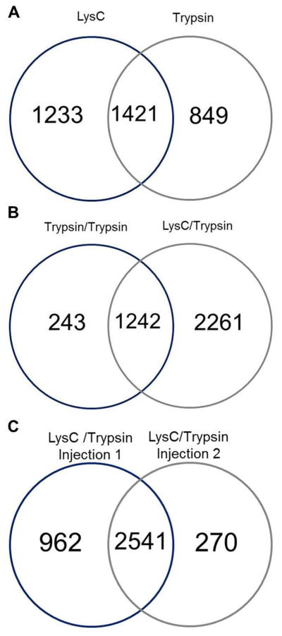
Figure 3.
Venn diagrams of proteins found in the proteome of P30 mouse cochlear sensory epithelium, when using multiple enzyme digestions. Number of proteins identified when performing a (A) first digestion with LysC and a second digestion with trypsin, (B) first digestion with trypsin or LysC followed by a second digestion with trypsin, and (C) LysC and trypsin digestion with multiple injections of each fraction.

