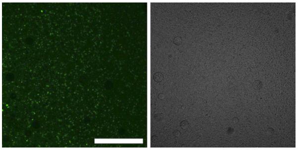
Figure 2.

Fluorescence (left) and light (right) microscopy of nano- and micro-droplets and their conglomerates in physiologic saline after centrifugation from fluorescein isothiocyanate-dextran solution; note that nano-droplets agglomerate and coalesce during centrifugation, which allows their optical visualization; large non-fluorescent circles are air bubbles. Scale bar = 0.2 mm.
