
Fig. 4.
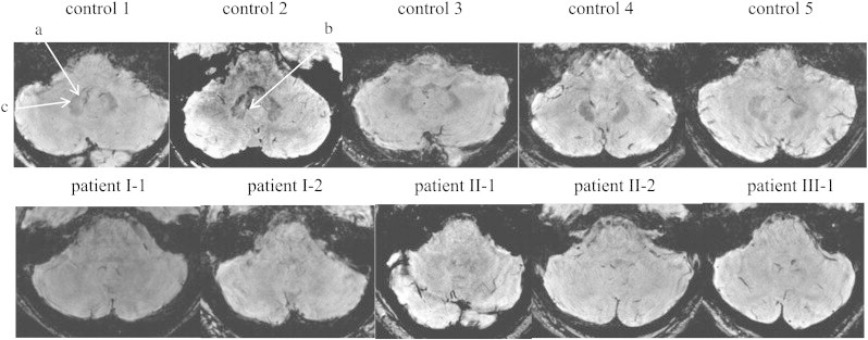
Representative axial slices of 3D fast field echo sequences of susceptibility-weighted images in controls (top raw) and in patients (bottom raw). a: Dentate nucleus; b: the hilus; c: toothed appearance after which the dentate nucleus is named.