
Fig. 1.
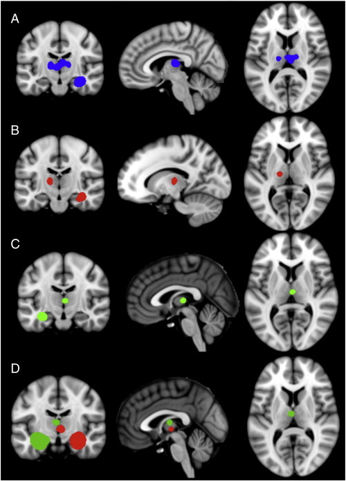
Significant areas of convergence in the VBM-ALE analysis in rectified (A), right medial temporal lobe epilepsy (B), and left medial temporal lobe epilepsy (C) groups. MACM analyses (D) of the left (green) and right (red) hippocampal tissue labels; note co-activation in the medial dorsal nucleus thalamus. All clusters thresholded at P < 0.05, corrected for multiple comparisons. See Supplementary materials for full montages of (A), (B) and (C).