
Figure 1.
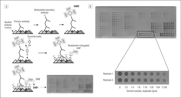
Reverse-phase protein microarray technology for quantitative analysis of human vitreous samples. A, Multiple vitreous samples and controls are immobilized on a nitrocellulose-coated slide and probed with a primary and secondary antibody, and the signal is amplified via horseradish peroxidase (HRP)–mediated deposition of biotinyl tyramide. B, The samples and controls are prepared in a 2-fold dilution series, allowing analysis within the linear dynamic range for each sample-antibody pair. DAB indicates diaminobenzidine complex; SABC, streptavidin-biotin; U, undiluted.