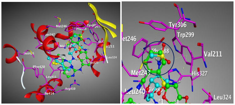
Figure 13. Comparison of the binding modes of compound 1 and compound 3 to PXR LBD.

Main interacting side chains identified through docking of compound 1 (green) and compound 3 (cyan) with the PXR LBD. The aryl group of compound 1 can form pi-pi interactions but not the methyl group of compound 3 shown as circle (blue).
