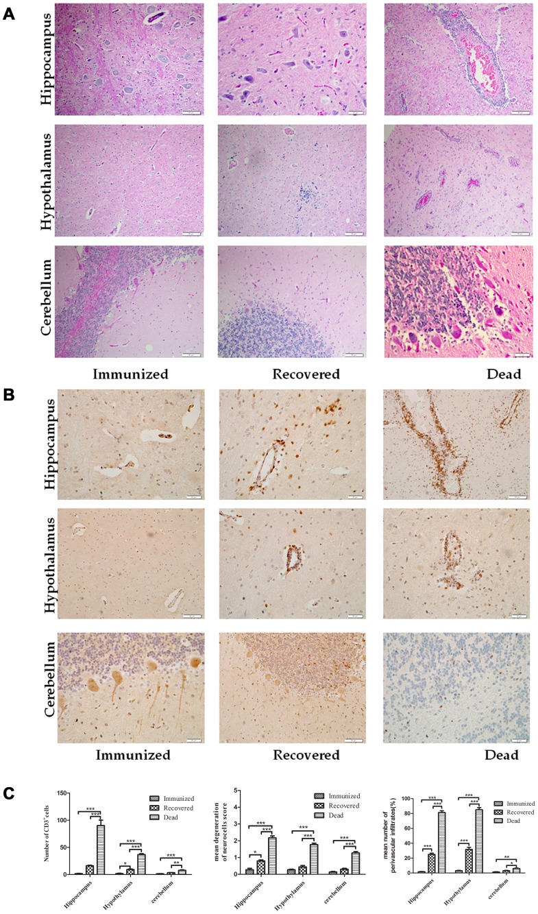
Figure 3. Infiltration of inflammatory cells into the CNS of dogs.
Paraffin sections obtained from DRV Mexico infected dog brains were subjected to HE staining (A) or immunohistochemistry for detecting CD3-positive cells (B). Three serial sections and two vessels were selected from each dog for quantification, and the average numbers of CD3-positive cells, degenerated neurons and perivascular infiltrated cells obtained were used for statistical analysis (C). Statistical analysis was performed by student T test and the symbol *** indicates a p value <0.0001, ** indicates a p value <0.001 and * indicates a p value <0.05.

