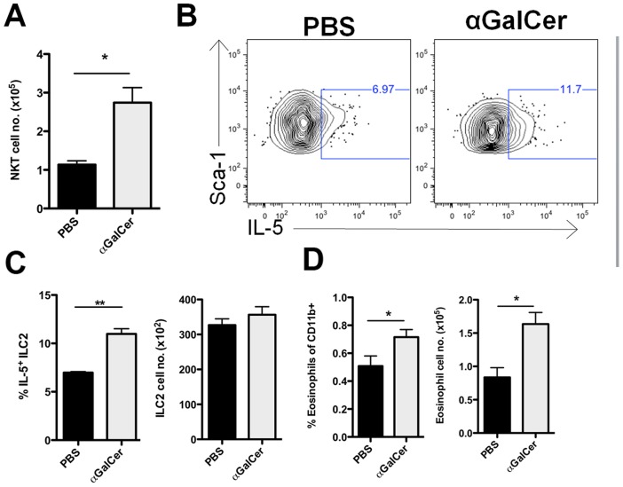
Figure 7. αGC administration increases both IL-5+ ILC2 and eosinophil numbers in the lungs.
(A) 1 µg/ml αGC was injected i.p. at 3 and 5 d.p.i.. Lungs were harvested at 7 d.p.i. and total NKT cells were quantified by use of CD3 and CD1d tetramer staining. (B) Representative FACS plots of intracellular IL-5 after 24 hours of ex vivo culture. (C) Frequency of IL-5+ ILC2 (left panel) and absolute number of ILC2 at 7 d.p.i. (right panel). (D) Frequency (left panel) and absolute number (right panel) of eosinophils detected in the lung at 7 d.p.i.. Data is representative of 2 independent experiments with 3 mice each. Bars = +/− SEM. *p<.05, **p<.01.

