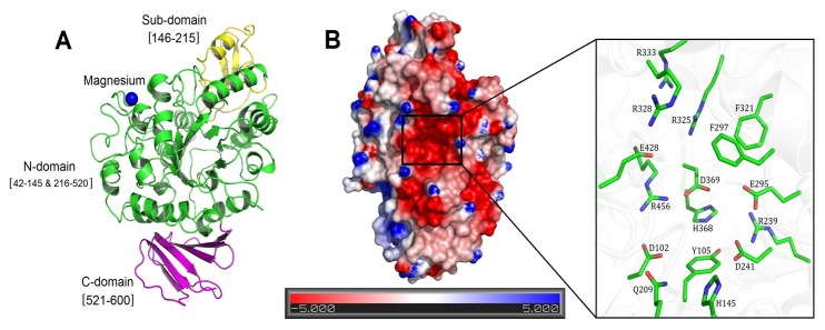
Figure 2. The crystal structure of native NX-5.

(A) The overall structure of native NX-5 protein. The N-domain, Sub-domain and the C-domain are colored in green, yellow and magenta, respectively. The magnesium ion is depicted as a blue sphere. (B) The charge state of the solvent-accessible surface of NX-5 is predominantly basic. The surface was colored according to the electrostatic potential of the molecule. A zoomed view of the key residues in the active site pocket is also shown.
