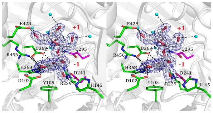
Figure 3. A stereo view of the binding mode between sucrose and NX-5 protein in the E295Q/sucrose complex.
The binding mode of sucrose in the E295Q/sucrose complex is shown in a stereoscopic view. The 2Fo-Fc electron density maps are contoured at 1σ. The water molecule is highlighted as a cyan ball and the hydrogen bonds are depicted as dashed lines.

