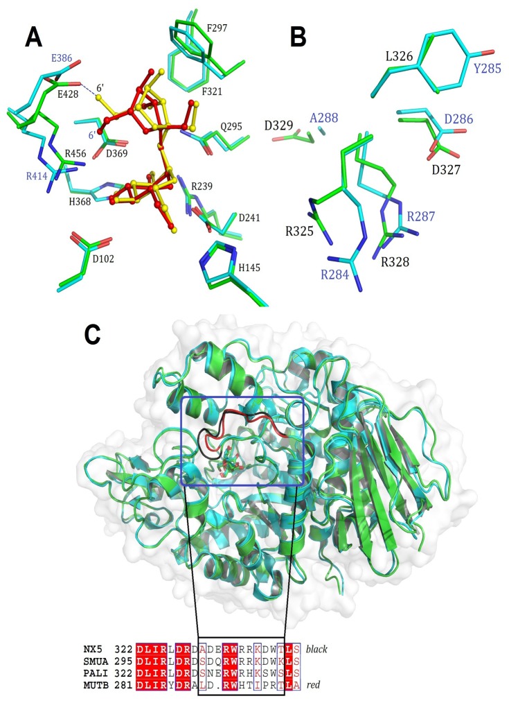
Figure 4. Structural difference between.
NX-5/sucrose and MutB/sucrose.
(A) Structural alignment of the active site residues between NX-5/sucrose (PDB ID: 4HPH, green) and MutB/sucrose (2PWE, cyan). The sucrose molecule in NX-5 is colored yellow, whereas that in MutB is colored red. Residues from NX-5 are labeled. (B) Structural comparison of325RLDRD329 in NX-5 (green) and284RYDRA288 (cyan) residues in MutB. (C) Structural and sequence comparison of loop330-339 (black) in NX-5 and the corresponding loop289-297 (red) of MutB.

