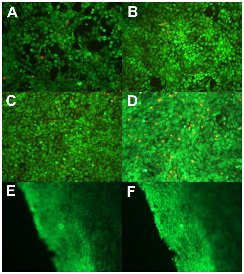
Figure 3. Live-dead assay of mMSC in hydrogels a continuous seven days in culture.
A, day 1; B, day 3; C, day 5; D, day 7. The living cells were stained with calcein AM (green) and the dead cells were stained with EthD-1 (red). The images of E and F showed top and side views of a 3D cell-gel construct at day 7, respectively.

