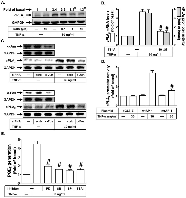
Figure 6. AP-1 is involved in TNF-α-induced cPLA2 expression.
(A) Cells were pretreated with Tanshinone IIA (TSIIA) for 1 h, and then incubated with TNF-α for 24 h. The protein levels of cPLA2 were determined by Western blot. (B) Cells were pretreated with Tanshinone IIA (TSIIA), and then incubated with TNF-α for 6 h. cPLA2 mRNA levels and promoter activity were determined. (C) Cells were transfected with scrambled, c-Jun, or c-Fos siRNA, and then incubated with TNF-α for 24 h. The protein levels of c-Jun, c-Fos, and cPLA2 were determined. (D) Cells were transfected with pGL3-empty, wild-type cPLA2 promoter, or AP-1-mutated cPLA2 promoter, and then incubated with TNF-α for 6 h. The promoter activity of cPLA2 was determined in the cell lysates. (E) Cells were pretreated with PD98059 (10 µM), SB202190 (10 µM), SP600125 (10 µM), or Tanshinone IIA (TSIIA; 10 µM) for 1 h, and then incubated with TNF-α for 24 h. The media were collected and analyzed for PGE2 release. Data are expressed as mean±S.E.M. of three independent experiments. # P<0.01, as compared with the cells exposed to TNF-α alone (A, B, and E). # P<0.01, as compared with cells transfected with wild-type cPLA2 promoter stimulated by TNF-α (D).

