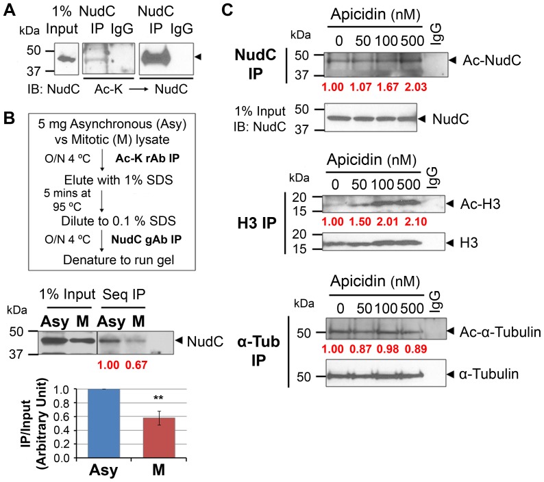
Figure 1. NudC is acetylated in interphase and deacetylated in mitosis.
(A) Unperturbed HeLa cell lysates (1 mg) were immunoprecipitated (IP) for NudC under denaturing conditions (used subsequently to examine NudC acetylation) and blotted with anti-acetyl lysine (Ac-K) antibody then reblotted for total NudC. (B) Sequential IP (Seq IP) flow chart. Lysates (5 mg) from asynchronouse (Asy) and mitotic (M) cells are IP'd first with anti-Ac-K antibody followed by IP for NudC then blotted for NudC. NudC acetylation is analyzed by ImageJ as a ratio of NudC IP over Input and normalized to R (fold change in red). Mean ± SEM of three independent experiments. **, p<0.02. (C) Cells were synchronized by a double thymidine block and release (see Figure 4A) then enriched in mitosis with nocodazole (Noc) with or without incubation with increasing concentrations of apicidin. Lysates (2 mg) were IP'd for NudC, histone H3 (H3), or α-tubulin (α-Tub) as in (A), and blotted with anti-Ac-K then reblotted for NudC, H3, or α-Tub, respectively. NudC, H3, and α-Tub acetylation are analyzed by ImageJ as a ratio of Ac-protein over either input (NudC) or protein IP (H3, α-Tub) and normalized to 0 nM Apicidin treatment (fold change in red). Data represent two to three independent experiments.

