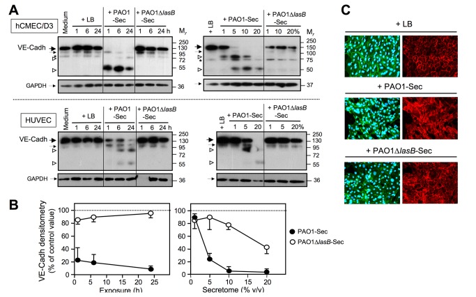
Figure 6. The LasB-containing secretome induces cleavage of VE-cadherin in endothelial cells in culture.
(A) Confluent EC cultures were exposed for 1 to 24 h to low FCS culture medium alone or with 10% of either LB, PAO1-Sec, or PAO1∆lasB-Sec (left-hand panels), or for 24 h to culture medium with LB (20%) or with the bacterial secretomes in the range of 1 to 20% (right-hand panels). Total proteins were extracted from residual adherent cells and SDS-PAGE/IB was applied to disulfide-reduced protein extracts (5 μg per well) for analysis of cell-associated VE-cadherin (VE-Cadh) using an anti-VE-cadherin pAb (1 μg/ml; upper panels). Membranes were then reprobed with an anti-GAPDH mAb (0.2 μg/ml; lower panels). Portions of the films corresponding to the location of the relevant molecular species are shown with location on the left-hand side of full-length VE-cadherin (large black arrow), truncated VE-cadherin detected in control cells, and GAPDH (small black arrows), and VE-cadherin fragments generated by PAO1-Sec (open arrowheads). An asterisk indicates a non-specific interaction routinely observed with hCMEC/D3 cell extracts. Results illustrated are each representative of three or five independent experiments that were performed on HUVECs or hCMEC/D3 cells, respectively. (B) Densitometric analysis of bands corresponding to the full-length VE-cadherin species as illustrated in (A) is reported as the mean percentage + SEM or - SEM of the value measured for control cell extracts exposed to LB. (C) Confluent hCMEC/D3 cell cultures were exposed for 24 h to low FCS culture medium with 10% LB, PAO1-Sec, or PAO1∆lasB-Sec (Immuno). fluorescence microscopy was performed as described in the legend to Figure 5, with adherent cells being permeabilized with Triton X-100 following fixation, and before sequential labeling with the pAb against VE-cadherin (5 μg/ml, right-hand panels), then with Alexa Fluor 488-phalloidin and with DAPI (left-hand panels). Representative areas (100x magnification) are shown for one of three similar experiments performed on either hCMEC/D3 cell or HUVECs. Left-hand and right-hand panels show the same area of the same culture chamber. Primary Ab replacement by a corresponding nonspecific IgG resulted in low diffuse background labeling (not illustrated).

