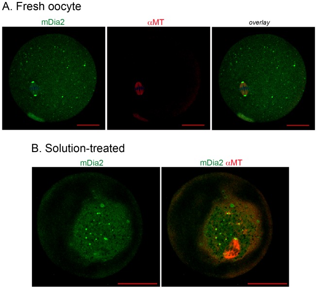
Figure 4. Co-staining of mDia2 and tubulin in MII oocytes and vitrification solution-treated oocytes.
A, Immunofluorescence staining of mDia2 and tubulin in fresh MII oocytes. mDia2, green; α-tubulin, red. B, Immunofluorescence staining of mDia2 and tubulin in oocytes treated with equilibration and vitrification solutions. Note the shrinkage of the ooplasm because of osmosis. mDia2, green; α-tubulin, red.

