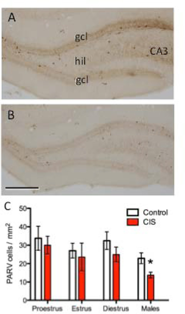
Figure 4. CIS decreases the number of PARV-labeled cells in the hilus of male, but not female, rats.
Representative photomicrographs show the distribution of PARV-labeled cells in the dentate gyrus from control (A) and CIS (B) male rats. CA3, cornu ammonis 3 region; gcl, granule cell layer; hil, hilus. Scale bar = 50 µm. C. The number of PARV-labeled cells was not significantly different in CIS and control female rats, regardless of estrous stage. Conversely, the number of PARV-labeled cells was significantly less (*p < 0.05) in CIS compared to control males. N = 6 rats per group.

