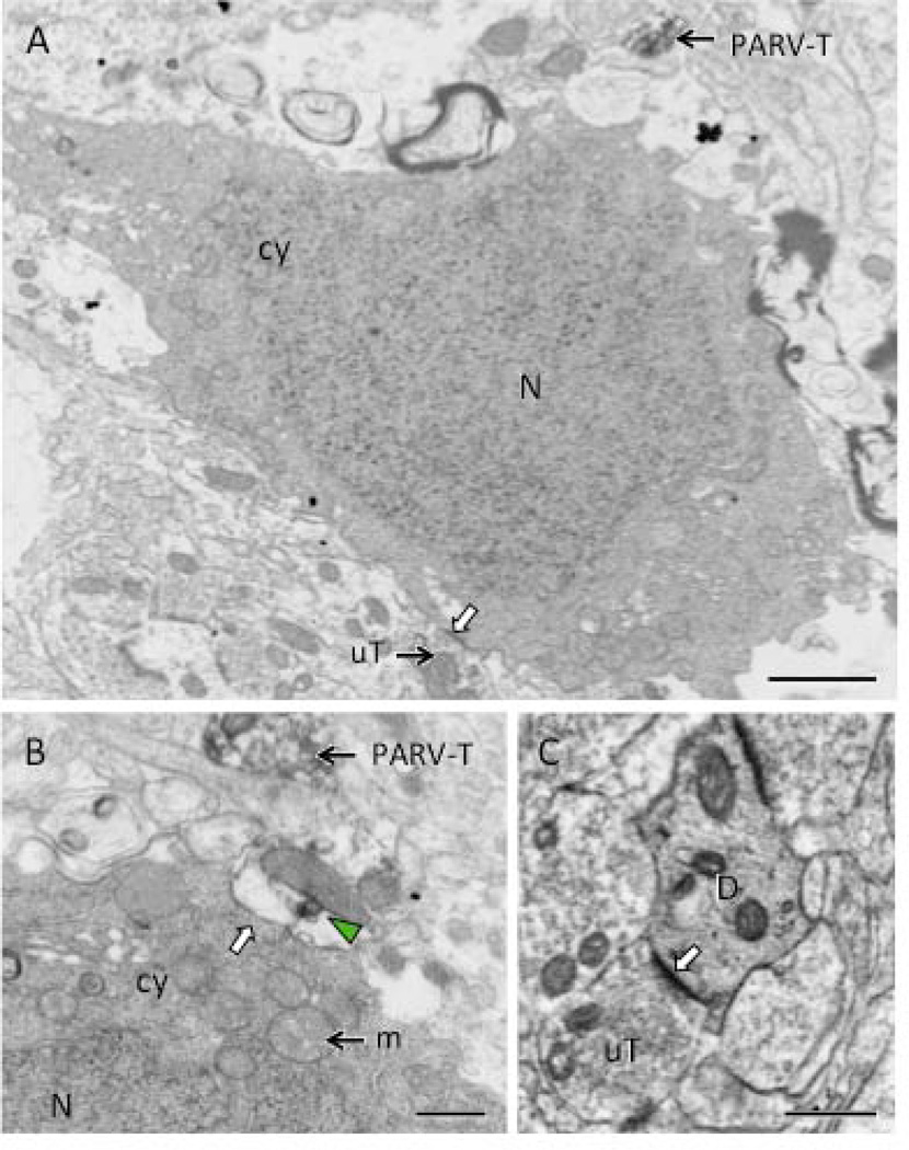
Figure 5. Electron microscopy reveals necrotic cell profiles in the hilus of male CIS rats.
A. A neuronal cell body, identified by its size and the presence of unlabeled terminals (uT) synapsing on the plasma membrane (white arrow), contains a dark nucleus (N) and dark cytoplasm (cy). A PARV-labeled terminal (PARV-T) is found nearby. B. At high magnification, a swollen profile with a patch of PARV-labeling (green arrowhead) abuts (white arrow) another neuron cell body with dark cytoplasm (cy) and nucleus (N). A PARV-labeled terminal (PARV-T) is found nearby. Numerous swollen mitochondria (m) are found in the cytoplasm of the profiles in A and B. C. A dendrite (D) with dark cytoplasm is contacted (white arrow) by an unlabeled terminal (uT). Scale bars = 500 nm.

