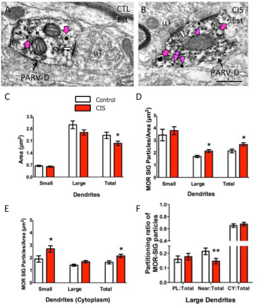
Figure 6. CIS affects the number and density of MOR SIG particles in PARV-labeled dendrites in female rats.
Examples of PARV-labeled dendrites (PARV-D) containing MOR SIG particles in the cytoplasm (magenta arrows), on the plasma membrane (magenta arrowhead) and near the plasma membrane (double magenta arrowheads) from control (A) and CIS (B) estrus females. Unlabeled terminals (uT) contact (white arrows) dual labeled dendrites. Scale bars = 500 nm. C. Following CIS, the area of total PARV-labeled dendrites was less (*p < 0.05) and tended to be less in large dendrites (p = 0.06) in CIS estrus females compared to controls. D. CIS significantly increased the total density of MOR SIG particles in large and total PARV-labeled dendrites of estrus females (*p < 0.05). E. CIS significantly increased (*p < 0.05) the cytoplasmic density of MOR SIG particles in small and total PARV-labeled dendrites and tended (p = 0.06) to increase the cytoplasmic density of MOR SIG particles in large PARV-labeled dendrites. F. CIS significantly decreased (**p < 0.01) the near plasma membrane MOR SIG particle distribution in of PARV-labeled dendrites in estrus females. N = 3 rats per group; n = 50 dendrites per rat.

