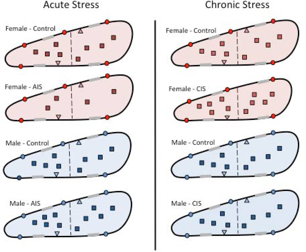
Figure 8. Schematic diagram summarizing the effects of acute and chronic stress on the density and trafficking of MORs in PARV dendrites in the hippocampus of female and male rats.
MORs are located on the plasmalemma (circle), near-plasmalemma (triangle) and in the cytoplasm (square) of PARV-containing dendrites in female and male rats. For ease of comparison, only estrus females are shown. Dashed line indicated division between small and large PARV-labeled dendrites and gray shadings on membranes indicate synapses. After AIS, fewer MORS are detected in the cytoplasm of small PARV-containing dendrites in females (red) whereas the proportion of MORs in cytoplasm compared to the total is greater in small and large PARV-containing dendrites in males (blue). After CIS, the numbers of PARV-labeled neurons decrease by about 30% in males but do not change in females (not shown). However, the size of large PARV-labeled dendrites decreases about 20% in females but not males. Following CIS, the density of MORs increases in the cytoplasm of small and large PARV labeled dendrites in females. Moreover, the proportion of near-plasmalemmal MORs compared to the total in large PARV-labeled dendrites decreases in females. After CIS, no changes in the density or trafficking of MORs are seen in the PARV-labeled dendrites in males.

