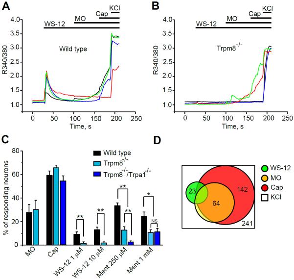
Fig. 4.
Effects of cooling agent WS-12 on mouse DRG neurons, measured by ratiometric Ca2+-imaging.
(A) Representative Ca2+-signals in wild-type DRG neurons elicited by WS-12 (1 μM), followed by mustard oil (MO, 70 μM), capsaicin (Cap, 1 μM) and KCl (40 mM).
(B) Representative Ca2+-signals in Trpm8−/− DRG neurons. Experiments performed as in (A).
(C) Percentage of wild-type, Trpm8−/− and Trpa1−/−/Trpm8−/−DRG neurons responding to Cap, MO, WS-12 (1 or 10 μM) and L-menthol (Ment, 250 μM or 1 mM). Each column shows average percentages from 5 to 8 separate tests and each test contains 40–60 neurons. Neurons were defined as responsive when the increase in Fura-2 emission ratio (340 nm/380 nm) in a given neuron exceeded 10% of the KCl response. **p <0.01, *p < 0.05, NS: no significance (p > 0.05).
(D) Population analysis of wild-type DRG neurons responding to WS-12, MO and Cap. Agonists were applied as in (A). (n = 241 neurons, 6 fields).

