Figure 4.
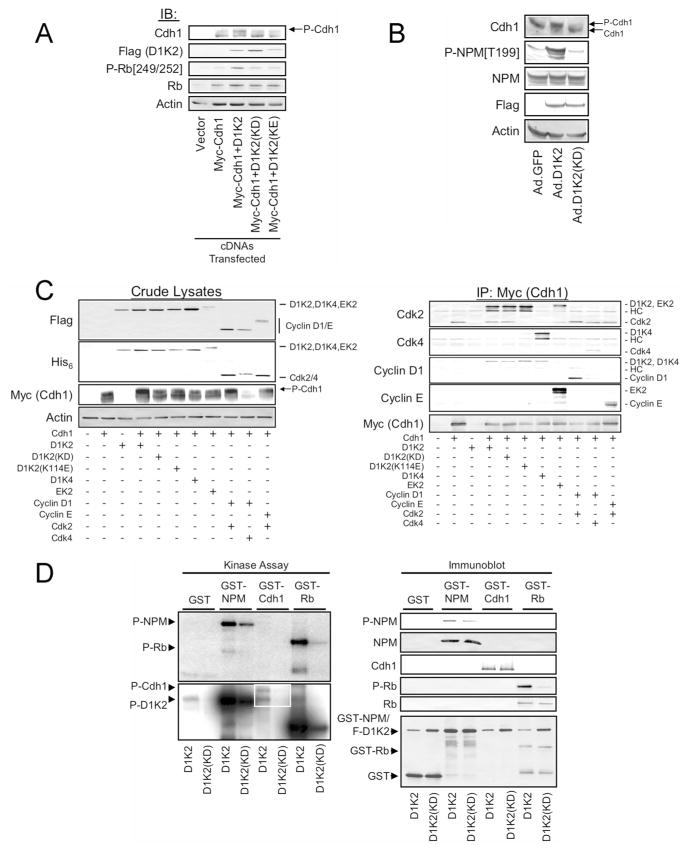
D1/K2 complexes and the Cyclin D1-Cdk2 fusion protein physically interact with and phosphorylate Cdh1. (A) Immunoblot analysis of cell lysates obtained from 293T cells transiently transfected with vectors encoding the indicated proteins. (B) Immunoblot analysis of cell lysates from NMuMG cells infected with adenoviruses encoding the indicated proteins. Flag staining denotes the presence of the D1K2 and D1K2(KD) proteins. (C) Immunoblot analysis of crude lysates (left panel) and affinity purified Cdh1 complexes (right panel) obtained from 293T cells transiently transfected with constructs encoding the indicated proteins. The identities of the bands are indicated to the right of some panels. (D) Autoradiograms of short (top left panel) and long (lower left panel) exposures and immunoblot analysis (right panel) of kinase assays using D1K2 and D1K2(KD) isolated from NMuMG cells infected with recombinant adenoviruses, and GST-NPM, GST-Cdh1, or GST-Rb as substrates, or GST alone as a control.

