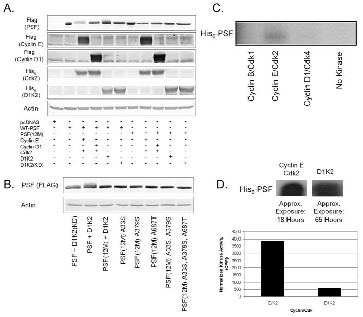
Figure 6.
Analysis of PSF phosphorylation. (A) Immunoblot analysis of FLAG-PSF or non-phosphorylatable PSF(12M) expressed with different Cyclin/Cdk complexes. (B) Immunoblot analysis of FLAG-PSF containing various mutations of potential phosphorylation sites expressed with D1K2. (C) Autoradiography of His6-PSF [γ-32P]-phosphorylated with different Cyclin/Cdk complexes. (D) Autoradiography of His6-PSF [γ-32P]-phosphorylated with either Cyclin E/Cdk2 or D1K2 (left panel) and quantitation of the same bands using a scintillation counter (right panel).

