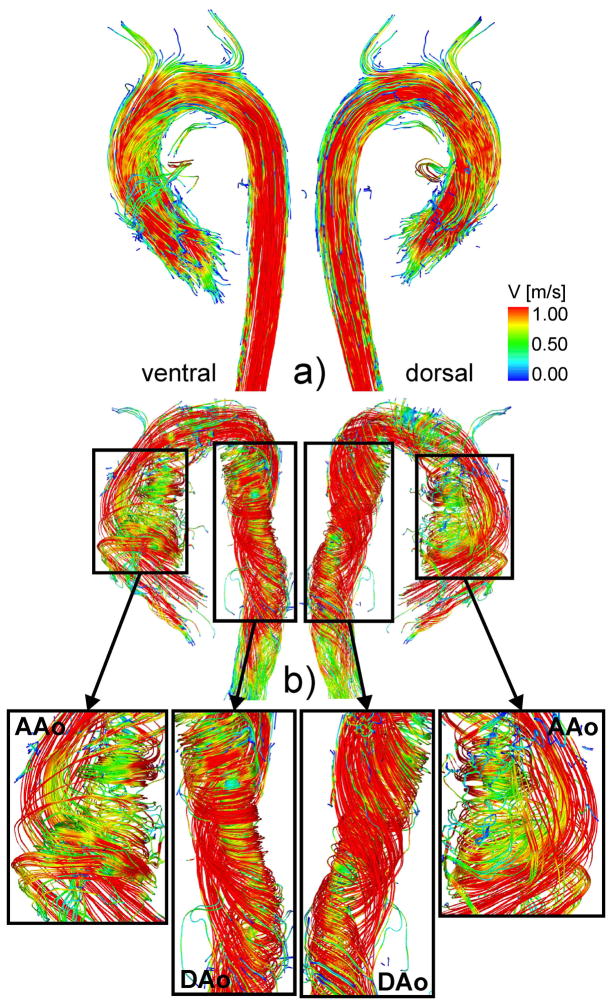
Fig. 1.
Comparison of streamlines and the formation of helical flow during peak systole.
top: Healthy subject shows helical flow as a normal feature in the aorta.
bottom: Patient with strong helical flow within the entire aorta.
The enlarged sections indicate various helical features of the flow pattern.

