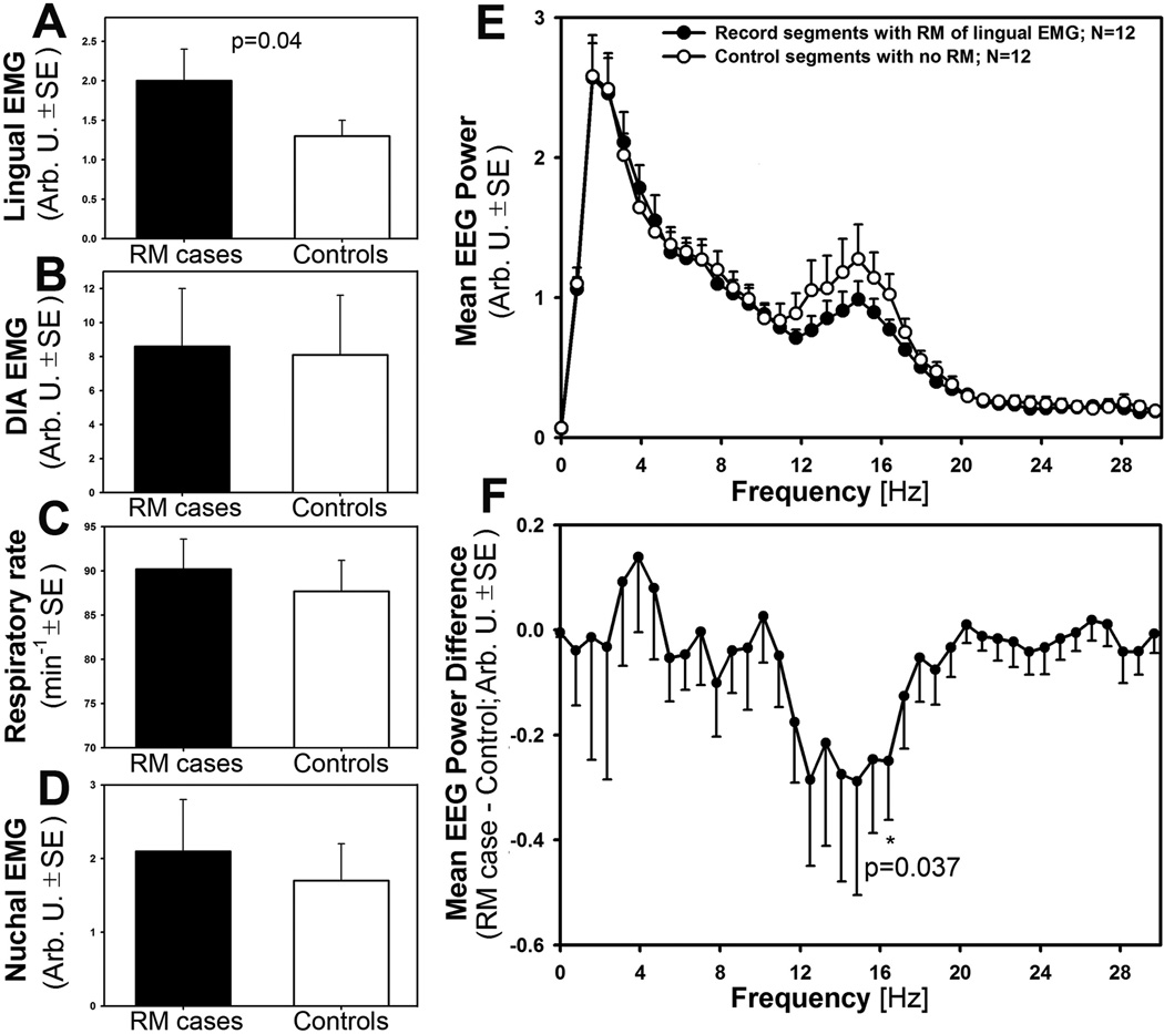
Figure 5.
The “case-control” analysis revealed that RM of lingual EMG is associated with deep SWS, as indicated by decreased cortical EEG power in a low beta-1 frequency band. In panels A–E, measures of lingual and diaphragmatic EMG, respiratory rate, postural tone, and EEG power are compared pairwise between adjacent episodes of SWS in which RM of lingual EMG occurred (RM cases) or was absent (Controls). As expected, lingual EMG amplitude was significantly higher in RM cases than in Controls (A), but other measures such as the amplitude of inspiratory modulation of diaphragmatic EMG (B), respiratory rate (C) and postural tone measured from nuchal EMG (D) did not differ. In contrast, the power spectra of cortical EEG differed between RM cases and Controls within one distinct frequency band (E). This band, 12.5–16.7 Hz, is designated as a low beta-1 or sigma in rats. F: expanded plot of the mean cortical EEG power difference between RM cases and Controls, with significant differences present at 16.7 Hz and across the entire 12.5–16.7 Hz range.

