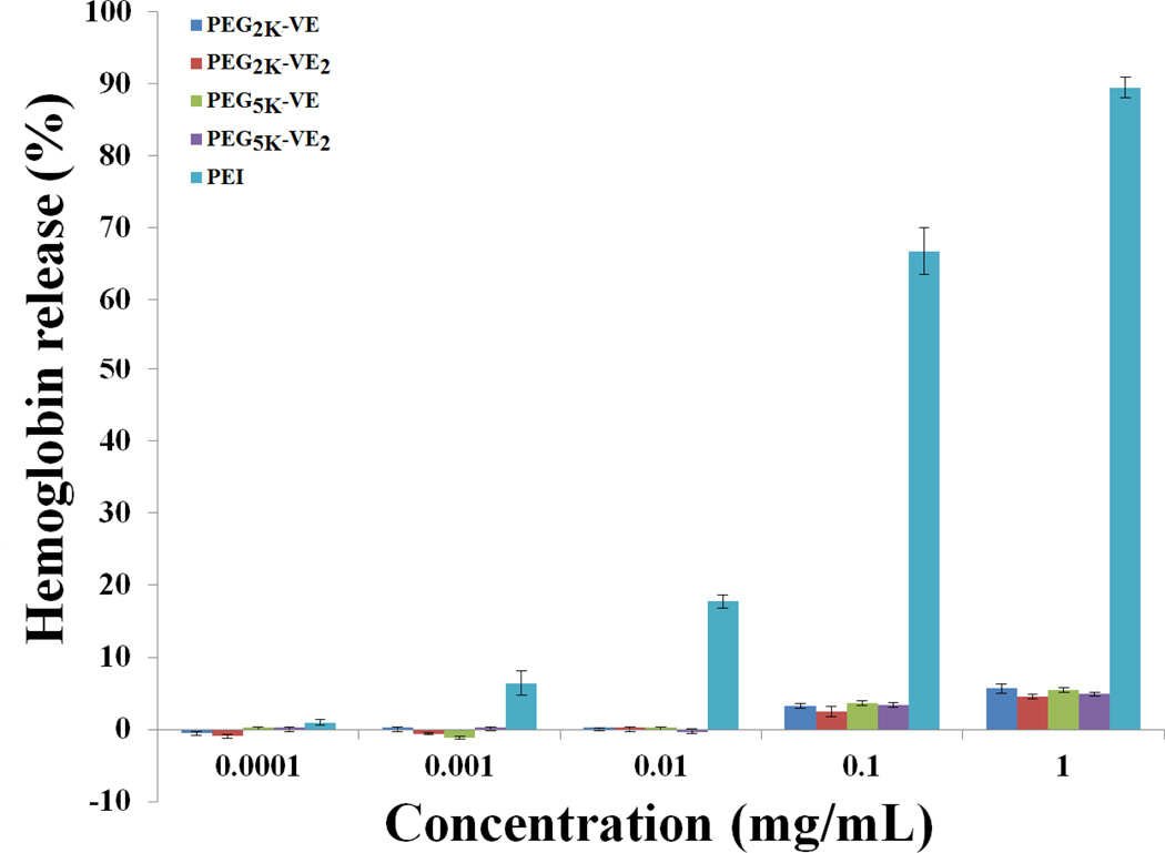
Fig. 5.
In vitro hemolysis assay of PEG-derivatized Vitamin E micelles compared with PEL PEG-VE micelles and PEI of various concentrations were incubated with rat red blood cells (RBCs) for 4 h at 37 °C in an incubator shaker. The degree of RBCs lysis was measured spectrophotometrically (λ=540 nm) according to the release of hemoglobin. 2% Triton X-100 and DPBS were used as a positive and negative control, respectively. Values reported are the means ± SD for triplicate samples.

