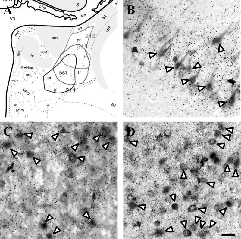
Figure 6.
Injection sites targeting the posterior subnuclei of the BST (A). Excitatory input to the BST arose from the vGluT1-positive vSub (B) and vGluT2-positive anterior PVT (C). A large number of FG-labeled cells were present in the medial CeA, most of which co-localized with GAD65 mRNA (D). White arrow heads indicate co-localization. Scale bar = 50 μm.

