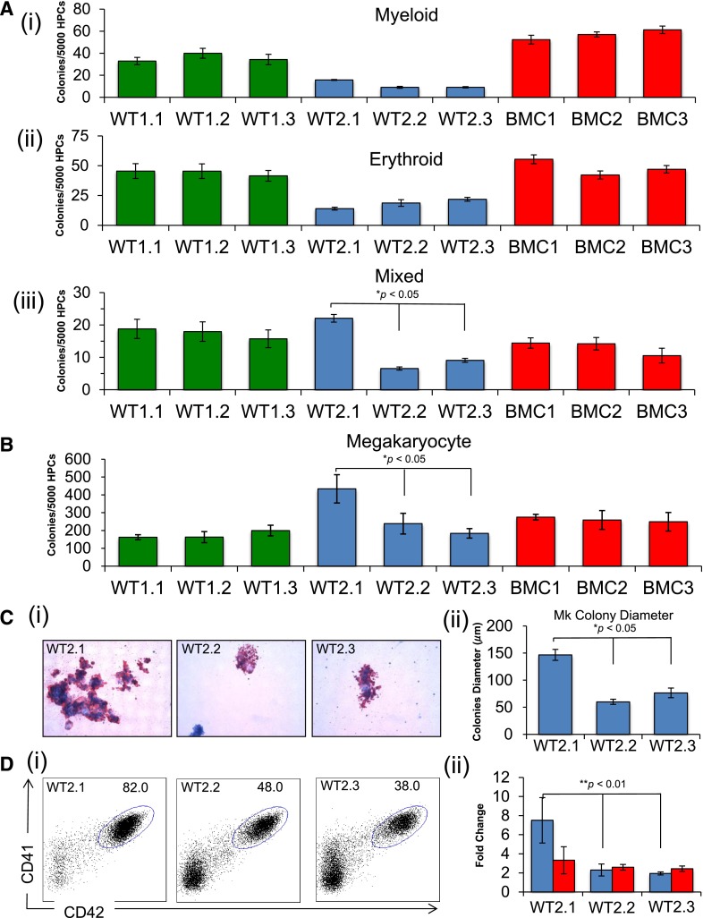
Figure 2.
In vitro differentiation of iPSC lines showing selective skewing toward megakaryopoiesis. (A) Methylcellulose progenitor assay of iPSC-derived HPCs from day 7 of differentiation. The panels show myeloid (i), erythroid (ii), and mixed (iii) colonies (mean ± SEM for 3 independent experiments, *P < .05). (B) Collagen-based colony assay to assess megakaryocyte (Mk) potential of iPSC-derived HPCs. (C) Images (i) of Mk colonies were captured by the Zeiss Axioskop2 microscope (Munich, Germany) (original magnification ×20), and (ii) size as a measure of Mk colony diameter (μm) (mean ± SEM for 3 independent experiments, *P < .05). (D) Assessment of megakaryocytic development from HPCs grown in liquid culture with thrombopoietin, interleukin 3, and SCF. The percentage of CD41a (αIIb, x-axis) and CD42a (GPIX, y-axis) Mk (i) are compared for CHOPWT2 lines, and (ii) total Mk number (blue) and non-Mk (red) cells are represented as fold change above the starting number of cells (mean ± SEM for 3 independent experiments, **P < .01).

