
Figure 6. Glycan fluorescent profiles from human AGP released by PNGase F and EndoS2.
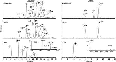
HILIC–FLD–MS of 2-AB-labelled glycans released from human AGP by PNGase F (left) and EndoS2 (right) respectively. 2-AB-labelled glycans were digested further with NAN1 and ABS, and subsequent bi-, tri- and tetra-antennary structures are indicated using the Oxford glycan nomenclature [45]. Ions were detected as [M−2H]2− (**) and [M−H]− (*) species.