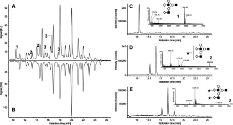
Figure 8. EndoS2 hydrolyses biantennary free glycans.

Bovine fetuin N-glycans were released with PNGase F, labelled with 2-AB, and analysed by HILIC–UHPLC–FLD–MS. Released N-glycans were digested further with EndoS2 to determine enzymatic activity on free glycans. Comparison of the fluorescent chromatograms of glycans after PNGase F (B) and subsequent EndoS2 digestion (A) identified three unique peaks (labelled 1, 2 and 3). These peaks correspond to three isomeric structures (A2G2, A2G2S1 and A2G2S2) and were detected primarily as m/z 1558.55 [M+H]+, 925.33 [M+2H]2+ and 1070.87 [M+2H]2+ ions respectively (C)–(E). Extracted ion chromatograms of A2G2S1 and A2G2S2 precursor ions identified structural isomers, presumably from variation in sialic acid linkages.
