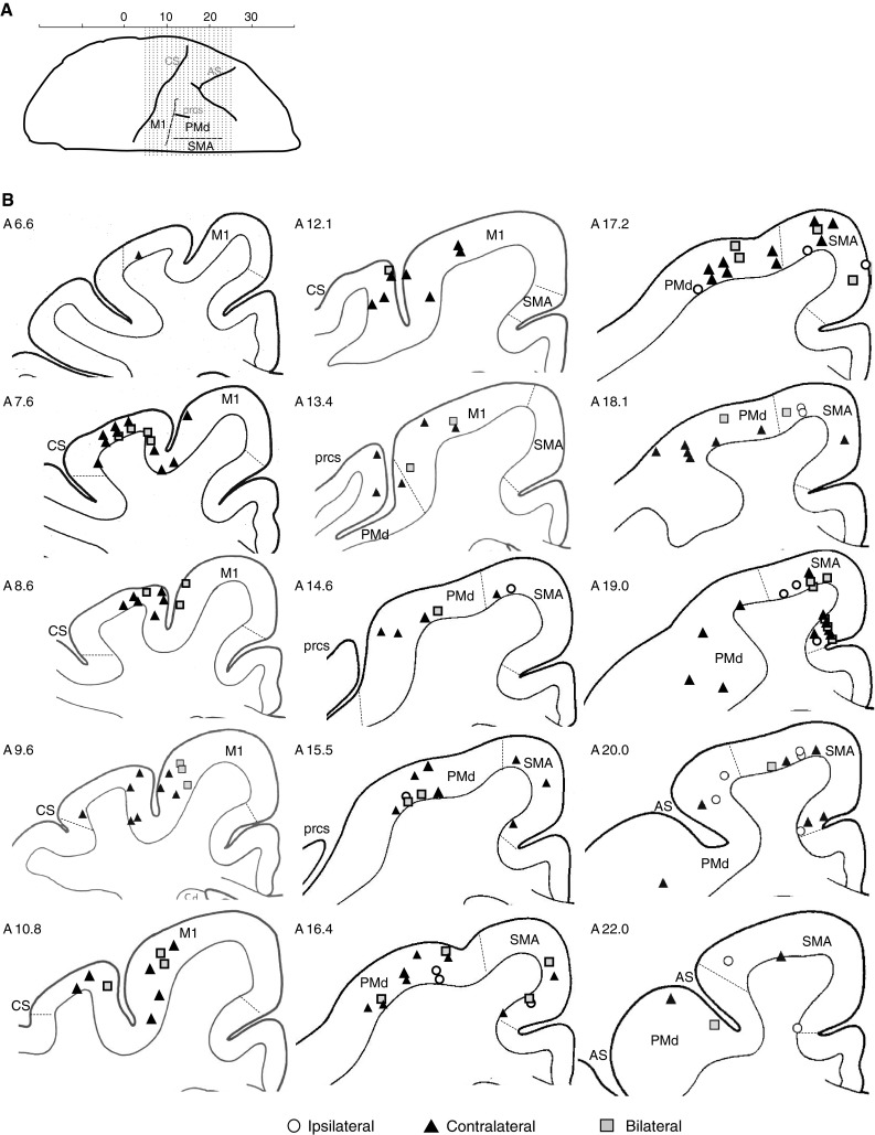
Fig. 2.

Schematic representation of the cerebral cortex illustrating responsive stimulation sites in each motor area. a A cranial view of the left cerebral cortex showing the position of M1, SMA, and PMd in relation to physical landmarks (from left to right is caudal to rostral and the lateral surface is uppermost). b The caudal to rostral representation of the location of cortical stimulation points that elicited EMG responses (cortical tracings based on Szabo and Cowan). White circles indicate ipsilateral response sites, black triangles indicate contralateral sites, and gray squares indicate bilateral sites. As shown, most of the ipsilateral sites were located in rostral and medial cortical areas (SMA). CS central sulcus, prcs precentral dimple, and AS arcuate sulcus
